AMYOTROPHIC LATERAL SCLEROSIS
-
Amyotrophic lateral sclerosis (ALS) is a neurodegenerative disease characterised by upper and lower motor neuron death with an ascending paralysis that eventually leads to death. The role of excitotoxicity is now recognised as an important factor in the ALS physiopathology.
GLUTAMATE and NMDA models
Glutamate receptors activation greatly contributes in mediating injury to motor neurons.
In in-vitro, a brief exposure to glutamate or NMDA causes neuronal death. This neuronal cell death is mainly due to the excessive stimulation of NMDA receptors.
-
Compound testing
Neuroprotectants are usually tested in this model but other treatments could also be considered. Please feel free to contact us to discuss the feasibility of your study.
-
Endpoints
☐ Neurite network
☐ Neuronal death
☐ Neuron survival
-
EXCITOTOXICITY on CORTICAL NEURONS
☐ GLUTAMATE testing - 13-day-old cortical neuron cultures are injured by an acute intoxication with glutamate.
The neuroprotective effect of compounds is evaluated based on their ability to inhibit on this damage. In the pre-treatment protocol, test compound is added 24h before intoxication and in the post-treatment one, test compound is added immediately after intoxication. Neuronal death is assessed by measuring LDH activity in the media at 24h after glutamate exposure.
- 13-day-old cortical neuron cultures are injured by an acute intoxication with glutamate.
-
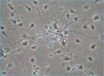
13-day old culture of cortical neuron on CONTROL condition. -
-
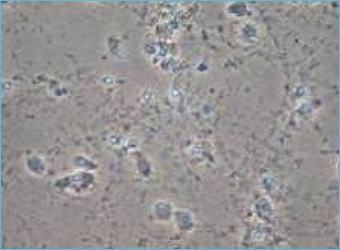
13-day old culture of cortical neuron injured by GLUTAMATE (10min, 75µM).
-
35,000 neurons per well
-
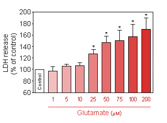
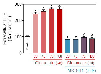
Release of LDH (CytoTox 96® Non-Radioactive Cytotoxicity Assay, Promega) in response to increasing doses of acute (10 min) glutamate intoxication on 13-day old culture of cortical neuron. -
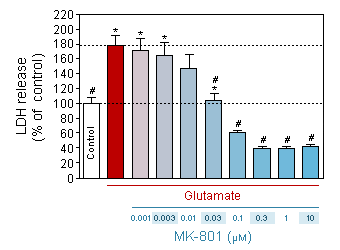
Effect of MK-801 on neuronal death induced by glutamate (75 µM, 10 min).
* p<0.05 compare the control group;
# p<0.05 compare the intoxicated group -
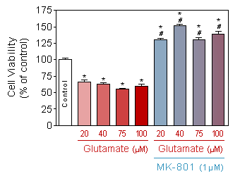
Evaluation of neuronal viability (Quantification of the presence of ATP by CellTiter Glo, Promega) in response to increasing doses of acute (10 min) glutamate intoxication on 13-day old culture of cortical neuron, in presence or absence of 1µM of MK-801.
* p<0.05 compare the control group;
# p<0.05 compare the corresponding glutamate group
-
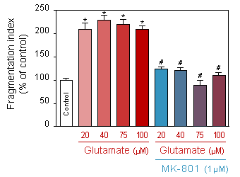
Evaluation of neuronal toxicity (quantification of binding of cell impermeant cyanine dye to dsDNA by CellTox Green, Promega) in response to increasing doses of acute (10 min) glutamate intoxication on 13-day old culture of cortical neuron, in presence or absence of 1µM of MK-801.
* p<0.05 compare the control group;
# p<0.05 compare the corresponding glutamate group -
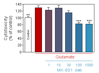
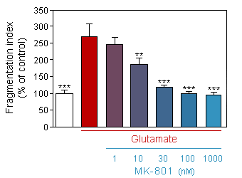
Evaluation of neuronal toxicity (quantification of binding of cell impermeant cyanine dye to dsDNA by CellTox Green, Promega) in response to acute (10 min) glutamate intoxication on 13-day old culture of cortical neuron, in presence or absence of increasing doses of MK-801.
* p<0.05 compare the corresponding glutamate group -
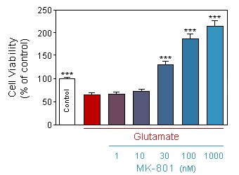
Evaluation of neuronal viability (Quantification of the presence of ATP by CellTiter Glo, Promega) in response to acute (10 min) glutamate intoxication on 13-day old culture of cortical neuron, in presence or absence of increasing doses of MK-801.
* p<0.05 compare the corresponding glutamate group
-
10,000 neurons per well
-
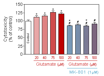
Release of LDH (CytoTox 96® Non-Radioactive Cytotoxicity Assay, Promega) in response to increasing doses of acute (10 min) glutamate intoxication on 13-day old culture of cortical neuron, in presence or absence of 1µM of MK-801.
* p<0.05 compare the control group;
# p<0.05 compare the corresponding glutamate group -
Effect of increased doses of acute (10min) glutamate on neurite network of 13-day old culture of cortical neuron, in presence or absence of 1µM of MK-801.
* p<0.05 compare the control group;
# p<0.05 compare the corresponding glutamate group -
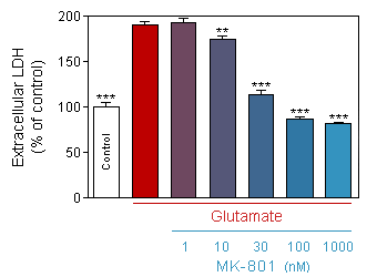
Release of LDH (CytoTox 96® Non-Radioactive Cytotoxicity Assay, Promega) in response to acute (10 min) glutamate intoxication on 13-day old culture of cortical neuron, in presence or absence of increasing doses of MK-801.
* p<0.05 compare the corresponding glutamate group
-
Effect of acute (10min) glutamate on neurite network of 13-day old culture of cortical neuron, in presence or absence of increasing doses of MK-801.
* p<0.05 compare the corresponding glutamate group -
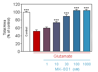
Effect of acute (10min) glutamate on total area of neurite network of 13-day old culture of cortical neuron, in presence or absence of increasing doses of MK-801.
* p<0.05 compare the corresponding glutamate group -
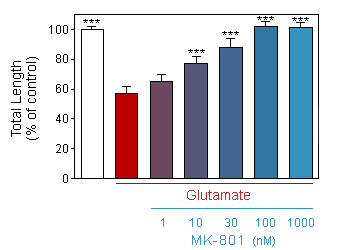
Effect of acute (10min) glutamate on total length of neurite of 13-day old culture of cortical neuron, in presence or absence of increasing doses of MK-801.
* p<0.05 compare the corresponding glutamate group
-
☐ NMDA testing - 12-day-old cortical neuron cultures are injured by an acute intoxication with NMDA. The neuroprotective effect of compounds is evaluated based on their ability to inhibit on this damage. In the pre-treatment protocol, test compound is added 24h before intoxication and in the post-treatment one, test compound is added immediately after intoxication. Neuronal death is assessed by measuring LDH activity in the media at 48h after NMDA exposure.
-
-
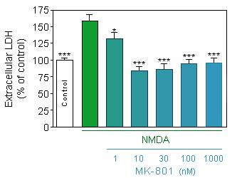
Release of LDH (CytoTox 96® Non-Radioactive Cytotoxicity Assay, Promega) in response to acute (75µM, 10 min) NMDA intoxication on 12-day old culture of cortical neuron at density of 10000 cells/well, in presence or absence of increasing doses of MK-801.
* p<0.05 compare the corresponding glutamate group -
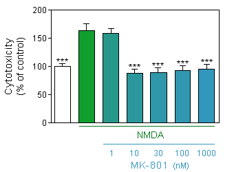
Evaluation of neuronal toxicity (quantification of binding of cell impermeant cyanine dye to dsDNA by CellTox Green, Promega) in response to acute (75µM, 10 min) NMDA intoxication on 12-day old culture of cortical neuron at density of 35000 cells/well, in presence or absence of increasing doses of MK-801.
* p<0.05 compare the corresponding glutamate group
-
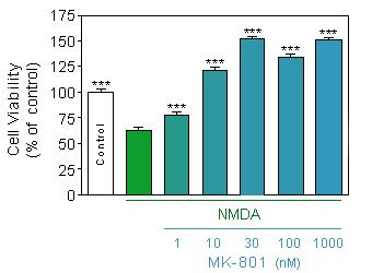
Evaluation of neuronal viability (Quantification of the presence of ATP by CellTiter Glo, Promega) in response to acute (75µM, 10 min) NMDA intoxication on 12-day old culture of cortical neuron at density of 35000 cells/well, in presence or absence of increasing doses of MK-801.
* p<0.05 compare the corresponding glutamate group -
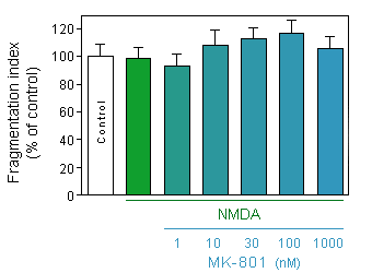
Effect of acute (75µM, 10min) NMDA on neurite network of 12-day old culture of cortical neuron at density of 10000 cells/well, in presence or absence of increasing doses of MK-801.
* p<0.05 compare the corresponding glutamate group
You could also be interested in
-
Co-culture
A functional model of motor unit RAT NERVE / HUMAN MUSCLE coculture.
-
Histomorphometry
Nerve function and axon morphology studies.

